|
|
English Structures 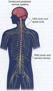 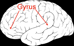 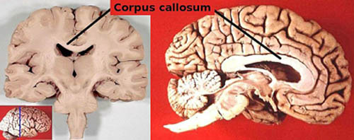 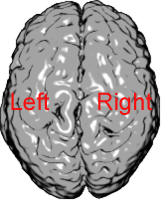 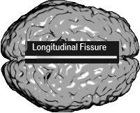 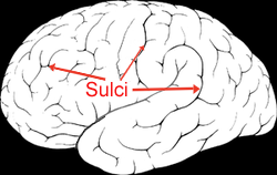 The brain stem (or brainstem) is the lower part of the brain, adjoining and structurally continuous with the spinal cord. It is one part of the brain included in the "reptilian brain" description. It is important for linking the brain to the peripheral nervous system. The brain stem also plays an important role in the regulation of cardiac and respiratory function. Respiratory control is a necessary component of speech production. The spinal cord carries out two main functions:
The spinal cord is partly involved in the contralateral control of the body. Sensory information from the left side of the body is processed in the right hemisphere of the brain, and vice versa. In some cases this crossing over occurs as soon as the impulses enter the spinal cord. In other cases, it does not take place until the brain itself has been reached. The nature of contralateral control has been a factor in research on the brain and learning how it processes language. The cerebellum is a region of the brain that plays an important role in the integration of sensory perception, coordination and motor control. In other words, it is involved in balance and movement. It does not play a role in the processing of language. The occipital lobe is the visual processing center of the mammalian brain containing most of the anatomical region of the visual cortex.The primary visual cortex is Brodmann area 17, commonly called V1 (visual one). Because of its role in processing visual information, this lobe is important in reading. The parietal lobe plays important roles in integrating sensory
information from various parts of the body, knowledge of numbers and their
relations and in the manipulation of objects. Portions of the parietal
lobe are involved with visuospatial processing. Much less is known about
this lobe than the other three in the cerebrum.
Language and the Brain
|
|||
|
Review: Language is a system of arbitrary symbols that humans use to create meaningful communication with other users of the same language. Human language differs from animal communication.
Why Humans Have Language
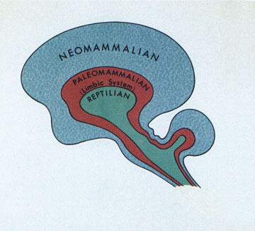
Anatomy of the Brain As described in Lesson 1 and mentioned in the review above, the human nervous sytem is directly tied to the ability of humans to use language. A closer study of the brain is in order, then, to understand the nature of human language. This lesson begins with a look at the general anatomy of the brain, the anatomy of the specific areas that process language and how the brain processes language, and finally discusses the ways that researchers have studied the brain to learn how it processes language. The brain is the part of the central nervous system located in the skull at the top end of the spinal cord. The brain and the spinal cord together make up the central nervous system. The peripheral nervous system connects the spinal cord and brain to the other organs in the body. See a diagram of both the Central Nervous System and the Peripheral Nervous System. The peripheral nervous system can be further divided, and so can the brain. Here we will look only at the parts of the brain. The Basic Building Blocks -- Neurons Neurons are the nerve cells that are the information processing blocks of the brain. There are about 100 billion neurons in the brain, which interconnect in complex networks. Each neuron can connect with about 10,000 other neurons. A neuron consists of a cell body with dendrite branches extending out from it (and a cell nucleus inside of it) and an axon. The axon extends out from the cell body opposite the dendrites and has its own branches called terminals. Click on Synapse to see a simulation of an electro-chemical message traveling through a neuron: the dendrites receive a message from another neuron, which then passes along the axon, and is sent out via the terminals to another neuron's dendrites. The space between one neuron's dendrites and another neuron's terminals is called a synapse. Click on Axon Growth to see a simulation of neuron growth forming a new connection between two cells. The dendrite of one cell first "sprouts" new branches that then extend out to the axon terminal of another neuron that is sending out a signal, thus making a new path for a chemical signal. When a person learns something, this process of developing a new path is what is actually happening in the brain. Dendrite growth to form a new connection requires time. The beginning of new growth that doesn't have enough time to make a new connection will be reabsorbed back into the old dendrite. The growth can be paused, but the new growth must continue to receive signals in order to be fully established.The "Triune" Brain One popular but simplistic description of the brain identifies three levels to the brain, which have evolved from the bottom up. Because of the three levels, it is called the "Triune Brain" description. The lowest level, the first to evolve, is referred to as "the Reptilian Brain." This level is responsible for basic life functions and is called reptilian to capture that fact. The middle level is referred to as "the Mammalian Brain" or "The Leopard Brain." This level includes the parts of the brain that house emotions, one of the features that makes mammals different from reptiles. The third and highest level, the last to evolve, is referred to as "the Primate Brain." Since humans are classified as primates, a brain with all three levels is the type of brain that humans have. All three levels of the brain are interconnected by neural networks.
While extremely simplistic, this description captures an important concept about the brain; the human brain is the most complex and highly evolved brain. Since language seems unique to humans, it must be in the part of the brain that humans have and other animals don't. That means language is located in the highest level, the cortex. What this overly simple view doesn't do, however, is explain how the cortex manages language, or why other primates don't have language like humans. Since Lesson 1 dealt with why other primates don't have language, this discussion will only continue with a focus on how the cortex manages language. A Scientific Description of the Brain The cortex of the brain is the most visible surface of the brain. It consists of a wrinkled mass that folds into itself like a crumpled up piece of paper. The inward folds are called sulci or fissures, and the outward folds are called gyri. It is divided into two hemispheres, corresponding to the right and left sides of the body. The two hemispheres are separated by a large fissure called the longitudinal fissure, but underneath the fissure is a mass that connects the two hemispheres: the corpus callosum.General Facts about the Cortex:
Activity: Features of the Cortex Click on the links in the definitions below to see diagrams and pictures of these features.Click on image to close. Sulcus/Sulci or fissures: the inward folds Gyrus/gyri: the outward folds making up the visible "surface." Hemispheres: The cortex is divided into two hemispheres, left and right each controlling different functions. Longitudinal Fissure: The longitudinal fissure is the large, deep sulcus separating the hemispheres. The Corpus Callosum: The corpus callosum is a bundle of nerve fibers that joins the hemispheres. Otherwise, the two hemispheres are completely separate from each other. Subdivisions of the CortexJust as the nervous sytem is divided and subdivided into smaller and smaller units, the cortex of the brain also is divided and subdivided according to its anatomy and its functions. We've already seen that the cortex is divided into hemispheres, and each hemisphere is divided into smaller sections called lobes. There are four major lobes in each hemisphere.
Activity: Functions of the Lobes Click and hold on each label for explanations and
definitions of those areas. The Frontal Lobe is the front part of the brain. It is involved in planning, organizing, problem solving, selective attention, personality and a variety of "higher cognitive functions&qout; including behavior and emotions. Broca's area is located in this lobe and is the area where syntactic language functions are carried out. The anterior (front) portion of the frontal lobe is called the prefrontal cortex. It is very important for the "higher cognitive functions" and the determination of the personality. The posterior (back) of the frontal lobe consists of the premotor and motor areas. Nerve cells that produce movement are located in the motor areas. The premotor areas serve to modify movements.
|
|||
